
A new approach for Ultra High Resolution Imaging in Biology
For years the RAITH CHIPSCANNER (https://www.raith.com/products/chipscanner.html) solutions have been used in semiconductor industry and intelligence service agencies. The tools originally developed for professionals dealing with integrated circuit design retrieval (?reverse engineering?) have now found a surprising application in imaging of biological tissues.
As reported in the September 2016 issue of Microscopy Today, Researchers of the George Washington University in the US applied RAITH CHIPSCANNER functionality to get an ultra-high resolution large area map of neuronal tissue.
Figure 1 shows a large area cross section of LgDel mouse spinal cord.
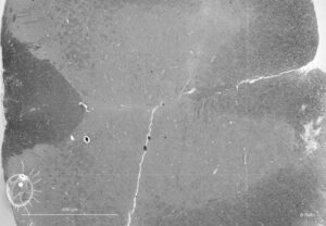
Figure 1: A large area cross section of spinal cord – George Washington University USA
This (lower resolution) picture however actually consists of many ultra-high resolution scanning electron microscope images seamlessly put together. Any detail of the spinal cord is readily available in ultra-high resolution. Figure 2 shows as an example a section of motor neurons of the same spinal cord.
The data the RAITH CHIPSCANNER captures from biological tissue could be very much used in the same way as satellite navigation internet tools: Viewing whole countries and zooming in at any point to see individual houses. In the case of the RAITH CHIPSCANNER data however this is possible with the precision of a few nano meters, there is nothing missing and there are no gaps.
The RAITH CHIPSCANNER originally developed to image integrated circuits helps now on better understanding of neuronal structure. In fact such a capability may play an important role in the context of the USA BRAIN initiative (https://www.whitehouse.gov/share/brain-initiative) or Human Brain Project in Europe (https://www.humanbrainproject.eu).
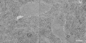
Figure 2: Section of motor neurons of the spinal cord from Figure 1 – George Washington University USA